Understanding The Laser Eye Assessment Process
The pre-operative assessment procedure
In order to safely alter the focus of the eye very detailed analysis of many parameters is necessary. On your visit to the clinic, your eyes will be measured on many machines.
None of these measurements are uncomfortable or dangerous and there is little or no risk involved in them.
An important measurement is the actual refractive state of your eye, i.e. what is your exact spectacle prescription. This must be measured with more than usual accuracy than would be the case just for glasses or contact lenses.
Another measurement is the mapping of both the front and back surfaces of the cornea. Also, the cornea must be sufficiently thick to allow the laser to safely operate. The eyes are also examined to exclude any other possible eye diseases which could affect the outcome.
Diseases such as keratoconus, where the shape of the cornea is abnormal, preclude laser surgery. The lens of the eye which sits just behind the pupil must also be healthy and the retina too must be free of significant disease. Most of these tests involve sitting in front of a machine and staring at a target.
They are not invasive and do not put the eye at risk. They yield highly important information to assess whether the eye is suitable for treatment and more importantly how much treatment is required.
Eye Assessment Machines
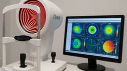
The Sirius Corneal Topographer
This device maps the anatomy of the front of the eye. It measures the shape of the cornea and looks for abnormal shapes or contours. It can analyse both the front surface and the back surface of the cornea. It can also analyse the shape of the interior of the front of the eye for those patients who might need an implant inside their eye to correct their vision.
Wavefront Analyser
This device measures how the eye handles light. It tracks 256 separate laser beams through the eye and sees where they pass. It then analyses the optical quality of the eye and gives very valuable information for surgical planning and assessing the success of the outcome.
OCT
Optical Coherence Tomography (OCT) is a device that allows us to examine the retina and the cornea in very considerable detail. It yields very valuable information regarding the morphology of these tissues. With it, we can see the rods and cones of the retina and assess their health. We can also analyse the anatomy of the optic nerve and make sure it is healthy.
Atlas
This is a device that maps the contours of the cornea. It gives valuable information in cases where there is a complicated spectacle prescription. In Presbyond surgery data from this machine is fed electronically into the laser as part of the surgical planning.
Optical Biometry
The technology is similar to OCT and it measures the length of the eye from the front of the cornea to the very back of the retina. It also measures other distances within the eye and gives information about the refracting power of the cornea.
Refraction
This is the examination that carefully measures exactly what spectacle prescription will put your eye into the best focus. It is essentially the same as a detailed spectacle examination with a good optician. In some patients, we also have to put drops into the eye to paralyse the focusing muscles for a short while. This allows more precise measurement in those cases.
This is the measurement that is used to calculate the exact treatment and as you can imagine is very important. It is more precise than ordinary glasses or contact lenses.
Pre-surgery Diagnostic Machines and Processes
| Eye Diagnostic Machine | Step | Description |
|---|---|---|
| Sirius Corneal Topographer | Consultation | Meet with a specialist to discuss the procedure, assess your candidacy, and address any questions or concerns. |
| Preparation | Remove any contact lenses and ensure your eyes are clean and free of debris before the scan. | |
| Positioning | Be comfortably seated in front of the Sirius Corneal Topographer machine. | |
| Scanning Process | Advanced imaging technology creates a detailed map of the cornea's surface. | |
| Analysis | Processed data generates a comprehensive visual representation of the cornea's topography. | |
| Review with Specialist | Discuss results with the specialist for surgical planning. | |
| Wavefront Analyser | Consultation | Meet with a specialist to discuss the procedure, assess candidacy, and address any questions or concerns. |
| Preparation | Remove contact lenses and ensure eyes are clean before the scan. | |
| Positioning | Be comfortably seated in front of the Wavefront Analyzer machine. | |
| Calibration | Machine is calibrated to focus on your eyes and collect precise measurements. | |
| Wavefront Analysis | Measures the entire optical system of your eyes. | |
| Data Processing | Processed data generates a detailed map of your eye's optical aberrations. | |
| OCT (Optical Coherence Tomography) | Consultation | Meet with a specialist to discuss the procedure, assess candidacy, and address any questions or concerns. |
| Preparation | No special preparation required before the scan. | |
| Positioning | Be comfortably seated in front of the OCT machine. | |
| Scan Process | Uses light waves to create cross-sectional images of the retina, providing detailed information about the eye's structure. | |
| Data Analysis | Processed images provide valuable insights for surgical planning. | |
| Atlas | Consultation | Meet with a specialist to discuss the procedure, assess candidacy, and address any questions or concerns. |
| Preparation | No special preparation required before the scan. | |
| Positioning | Be comfortably seated in front of the Atlas machine. | |
| Scan Process | Creates detailed measurements of the curvature and structure of the cornea. | |
| Data Analysis | Processed data aids in surgical planning and customizing treatment. | |
| Optical Biometry | Consultation | Meet with a specialist to discuss the procedure, assess candidacy, and address any questions or concerns. |
| Preparation | No special preparation required before the scan. | |
| Positioning | Be comfortably seated in front of the Optical Biometry machine. | |
| Measurement | Uses light to measure various parameters of the eye, including axial length and corneal curvature. | |
| Data Analysis | Processed measurements provide crucial information for accurate intraocular lens selection. | |
| Refraction | Consultation | Meet with a specialist to discuss the procedure, assess candidacy, and address any questions or concerns. |
| Preparation | No special preparation required. | |
| Positioning | Be comfortably seated in front of the Refraction machine. | |
| Testing | Assesses the refractive error of the eye to determine the appropriate correction. | |
| Prescription | Provides the precise prescription for glasses or contacts if needed post-surgery. |
Expected Times Of Each Process
Using Technology To Enhance Your Life
The diagnostic eye screening machines utilised at My-iClinic play a pivotal role in customising laser eye surgery treatments for each patient. The Sirius Corneal Topographer, Wavefront Analyzer, OCT, Atlas, Optical Biometry, and Refraction machines work collectively to provide a comprehensive understanding of the patient's eye structure and condition. Through precise measurements and detailed topographical maps, these machines enable our specialists to design highly personalised treatment plans. This level of customisation ensures that patients receive laser eye surgery that is tailored to their unique needs and delivers optimal outcomes.
As a result, patients can expect a transformative experience. The accuracy and specificity achieved through these diagnostic machines lead to enhanced surgical precision and post-operative visual acuity. With reduced reliance on glasses or contact lenses, patients regain newfound freedom and clarity in their vision. This personalised approach not only maximises the effectiveness of the surgery but also minimises any potential discomfort or complications. By leveraging state-of-the-art vision correction technology, My-iClinic empowers patients to embark on a journey towards improved vision, ultimately enhancing their quality of life.
Start Your Journey With My-iClinic
Experience the Clarity You Deserve!
Ready to free yourself from glasses and contacts? Start your journey to a clear vision today with My-iClinic. Schedule your consultation now and take the first step towards a brighter, lens-free future!
Find out more by Speaking to our team


