
In recent years, the field of ophthalmology has witnessed remarkable advancements in diagnostic eye equipment, revolutionising the landscape of vision correction. Cutting-edge technologies now enable eye care professionals to conduct highly precise and comprehensive assessments, facilitating personalised treatment plans for individuals seeking vision correction. One notable innovation is the integration of wavefront technology in diagnostic devices, allowing for a detailed analysis of the entire optical system of the eye. This technology provides a more nuanced understanding of aberrations and imperfections in the eye's optics, enabling surgeons to tailor laser vision correction procedures, such as LASIK or ReLEx Smile, with unprecedented accuracy.
Moreover, the advent of topography-guided diagnostics has elevated the precision of vision correction procedures to new heights. This technology creates detailed maps of the cornea's surface, aiding in the identification and correction of irregularities that may have previously gone undetected. These advancements not only enhance the overall safety and efficacy of vision correction surgeries but also contribute to the expanding scope of treatable visual conditions. As modern diagnostic eye equipment continues to evolve, the ability to diagnose and address even subtle optical anomalies becomes increasingly sophisticated, promising a future where personalised and precise vision correction is the norm rather than the exception.
Throughout this article, we will take a look at modern eye-screening and scanning technologies and look at how they can help with vision correction.
Visual Fields
Visual field testing is a diagnostic procedure used to assess the full horizontal and vertical range of what an individual can see, both centrally and peripherally. This test is crucial for detecting abnormalities in the visual field that may indicate various eye diseases or neurological conditions affecting the visual pathway. There are several methods and devices used for visual field testing, but the primary goal is to map out any areas of reduced sensitivity or complete visual loss.
One common method is the confrontation visual field test, where the examiner covers one eye while the patient focuses on an object with the uncovered eye. The examiner then moves an object, typically their fingers, from the periphery towards the centre of the visual field, and the patient signals when they first see it. This process is repeated in different areas of the visual field to create a map of the patient's peripheral vision.
More advanced and widely used methods involve automated perimetry, which uses specialised machines to systematically test the visual field. The patient looks at a fixed point while responding to the appearance of stimuli in their peripheral vision. This data is then analysed to generate a detailed visual field map, helping to identify specific patterns or defects that may be indicative of conditions like glaucoma, retinal diseases, or neurological disorders.
Visual field testing is an integral part of comprehensive eye exams, especially for individuals at risk of conditions affecting peripheral vision. Regular testing is essential for early detection and management of eye diseases, as it allows healthcare professionals to intervene before significant vision loss occurs.

iTrace
The iTrace system is an advanced diagnostic tool used for comprehensive assessment of the optical and visual characteristics of the eye. It provides valuable insights into various aspects of ocular function. The iTrace system combines wavefront aberrometry, corneal topography, and autorefraction to create a detailed analysis of the eye's optical system.
One of the key features of the iTrace system is its ability to measure and analyse the total visual function, including higher-order aberrations. Higher-order aberrations are subtle imperfections in the optical system of the eye that may contribute to visual disturbances such as glare, halos, and decreased contrast sensitivity. By capturing and evaluating these aberrations, the iTrace system helps eye care professionals design personalised treatment plans for vision correction procedures like LASIK or cataract surgery.
While not a substitute for traditional visual field testing, the iTrace system plays a vital role in providing a comprehensive understanding of the eye's optical quality. It assists in optimising the outcomes of vision correction surgeries by tailoring procedures to address not only refractive errors but also subtle aberrations that may impact visual acuity and quality. The iTrace's contribution to the field of eye care lies in its ability to enhance the precision and customisation of vision correction procedures, ultimately leading to improved patient outcomes and satisfaction.

IOL Master
The IOL Master, or Intraocular Lens Master, is a sophisticated diagnostic device primarily used in ophthalmology for precise measurements related to cataract surgery and intraocular lens (IOL) implantation. While not designed for visual field testing, the IOL Master serves a critical role in the preoperative planning process for cataract surgery.
The main function of the IOL Master is to measure the axial length of the eye, which is crucial for calculating the power of the intraocular lens that will be implanted during cataract surgery. Accurate measurements of axial length help ensure that the implanted lens will provide the appropriate refractive power, allowing for improved postoperative visual outcomes. Additionally, the IOL Master can measure other parameters, such as corneal curvature and anterior chamber depth, contributing to a more comprehensive understanding of the eye's anatomy.
By providing precise biometric data, the IOL Master aids surgeons in selecting the most suitable intraocular lens power for each patient, reducing the likelihood of postoperative refractive errors. This advanced technology has significantly improved the predictability and accuracy of cataract surgery outcomes, leading to enhanced visual results and patient satisfaction.
In summary, while the IOL Master is not used for visual field testing, its role in cataract surgery preparation is indispensable. It exemplifies the integration of technology in ophthalmology to optimise surgical planning and improve the overall quality of vision for patients undergoing cataract procedures.

Optical Coherence Tomography (OCT)
Optical Coherence Tomography (OCT) is a non-invasive imaging technology that plays a crucial role in diagnosing and managing various eye conditions. Unlike visual field testing, OCT provides detailed cross-sectional images of the eye's structures, allowing for a comprehensive analysis of the retina, optic nerve, and other ocular tissues.
Here's how OCT is used:
OCT is particularly valuable in the diagnosis and monitoring of retinal conditions, such as macular degeneration, diabetic retinopathy, and glaucoma. The device employs light waves to create high-resolution, cross-sectional images of the retina, enabling healthcare professionals to visualise the thickness and integrity of retinal layers. This information is vital for the early detection of structural changes or abnormalities in the eye, facilitating prompt intervention and management.
Additionally, OCT is widely used in the assessment of the optic nerve head and nerve fibre layer, crucial for the diagnosis and management of glaucoma. By providing detailed images of these structures, OCT aids in the early detection of glaucomatous damage, allowing for timely intervention to preserve vision.
Moreover, OCT is an advanced imaging technology that enhances the diagnosis and management of various eye conditions by providing detailed cross-sectional images of ocular structures. Its role is particularly significant in monitoring retinal health and assessing the optic nerve, contributing to improved patient care and outcomes in ophthalmology.

Sirius
The Sirius system is a versatile diagnostic platform in ophthalmology that integrates multiple imaging technologies to provide a comprehensive assessment of the anterior and posterior segments of the eye. Unlike visual field testing, the Sirius system focuses on corneal and anterior segment analysis, making it particularly valuable in the evaluation of conditions related to the front part of the eye. Here's how Sirius is utilised:
Corneal Topography
The Sirius system excels in corneal topography, offering detailed mapping of the corneal surface. This is invaluable for assessing corneal irregularities, astigmatism, and other refractive errors. The precise corneal measurements obtained with Sirius are essential for surgical planning, such as in procedures like LASIK or other corneal surgeries.
Anterior Segment Imaging
Sirius provides high-resolution images of the anterior segment, including the cornea, iris, and lens. This capability is crucial for diagnosing and managing conditions like corneal dystrophies, cataracts, and anterior segment tumours.
Pachymetry
The system includes pachymetry, a measurement of corneal thickness. This is essential for evaluating conditions such as keratoconus and glaucoma, where corneal thickness can be a significant factor.
Posterior Segment Imaging (optional)
Some models of the Sirius system may include modules for posterior segment imaging, allowing for the assessment of the retina and optic nerve head. This can be beneficial for comprehensive eye examinations.
Additionally, the Sirius system is a powerful diagnostic tool that combines corneal topography, anterior segment imaging, and optional posterior segment imaging. Its applications range from preoperative planning for refractive surgeries to the evaluation of corneal and anterior segment disorders, contributing to more precise diagnosis and treatment in ophthalmology.

Atlas
The Atlas Corneal Topography System is a diagnostic device used in ophthalmology to map the surface of the cornea. It provides detailed information about the shape, curvature, and thickness of the cornea. The device is particularly useful in assessing conditions such as corneal irregularities, astigmatism, and for preoperative planning of refractive surgeries like LASIK.
Key features of the Atlas Corneal Topography System may include:
Corneal Mapping: The device generates detailed maps of the cornea, allowing eye care professionals to analyse its topography and detect abnormalities.
Astigmatism Analysis: It helps in the precise measurement and analysis of astigmatism, aiding in the selection of appropriate treatments or surgical interventions.
Contact Lens Fitting: The system assists in fitting contact lenses by providing accurate measurements of the corneal surface.
Corneal Thickness Measurement: Some models may include pachymetry, measuring the thickness of the cornea, which is crucial for conditions like glaucoma.
The Atlas Corneal Topography System device contributes to the accurate diagnosis and management of corneal and refractive disorders, enhancing the overall quality of eye care.

Fundus
In ophthalmology, the term "Fundus" typically refers to the posterior part of the eye, including the retina, optic disc, and blood vessels. The examination of the fundus is crucial for assessing the health of the eye and diagnosing various ocular conditions. Here's an overview:
Fundus Examination
A fundus examination involves the evaluation of the back of the eye, which is illuminated and viewed through the pupil. Key components of the fundus examination include:
- The retina is the light-sensitive tissue lining the back of the eye. A fundus examination allows the assessment of the retina for signs of diseases such as diabetic retinopathy, age-related macular degeneration, and retinal detachments.
- The optic disc is where the optic nerve exits the eye. Its appearance is examined for signs of conditions like glaucoma.
- The blood vessels of the retina can be observed for abnormalities, including signs of hypertensive retinopathy or vascular diseases.
Technologies for Fundus Imaging
Several technologies are used to capture detailed images of the fundus:
- High-resolution images of the fundus are captured using specialised cameras. These images help document and track changes over time.
- Fluorescein Angiography involves the injection of a fluorescent dye into the bloodstream, which helps visualise the blood vessels in the retina. It is especially useful in diagnosing vascular abnormalities.
- Optical Coherence Tomography (OCT) is also used for detailed cross-sectional imaging of the retina, providing information about its thickness and structure.
Importance of Fundus Examination
Fundus examination is a cornerstone in the diagnosis and management of various eye diseases. It allows eye care professionals to detect and monitor conditions affecting the back of the eye, contributing to early intervention and better outcomes for patients. Regular fundus examinations are particularly crucial for individuals with conditions such as diabetes or a family history of eye diseases.
Moreover, the fundus examination is a fundamental aspect of eye care, providing valuable insights into the health of the retina and optic nerve. Advanced imaging technologies enhance the ability to diagnose and manage a wide range of ocular conditions.

iCare Tonometer
The iCare tonometer is a device used for measuring intraocular pressure (IOP), which is the pressure inside the eye. Monitoring intraocular pressure is essential in the management and diagnosis of various eye conditions, especially glaucoma.
Here is what to expect and why they are used:
Contactless Measurement
One notable feature of the iCare tonometer is its contactless measurement method. Unlike traditional tonometers that require anaesthetic eye drops and direct contact with the cornea, the iCare tonometer measures intraocular pressure with a small probe that barely touches the cornea's surface. This can make the measurement process more comfortable for the patient.
Quick and Painless
The measurement process with the iCare tonometer is quick and generally considered painless. This is advantageous in obtaining reliable pressure readings without causing discomfort to the patient.
IOP Monitoring in Glaucoma
Measuring intraocular pressure is crucial in the diagnosis and management of glaucoma, a group of eye conditions that can lead to optic nerve damage and vision loss. Elevated intraocular pressure is a significant risk factor for glaucoma, and regular monitoring helps in assessing disease progression and the effectiveness of treatment.
Importance of IOP Measurement
Glaucoma Detection: Elevated intraocular pressure is a major risk factor for glaucoma. Monitoring IOP helps in the early detection of glaucoma and allows for timely intervention to prevent vision loss.
Treatment Evaluation
For individuals with glaucoma or ocular hypertension, measuring intraocular pressure is crucial in evaluating the effectiveness of treatment, such as eye drops or surgery.
Baseline Measurement
Baseline intraocular pressure measurements are often taken during routine eye examinations. This establishes a reference point for future comparisons and aids in identifying any significant changes over time.
In summary, the iCare tonometer is a valuable tool for measuring intraocular pressure, contributing to the diagnosis and management of glaucoma and other eye conditions. Its contactless and user-friendly features enhance the patient experience during IOP monitoring. Regular intraocular pressure measurements are an integral part of comprehensive eye care, particularly for individuals at risk of or diagnosed with glaucoma.

Slit lamp
The slit lamp is a fundamental tool used in ophthalmology for a detailed examination of the anterior segment of the eye. It provides a highly magnified, three-dimensional view of the eye's structures, allowing eye care professionals to diagnose and monitor various eye conditions.
Slit Lamp Examination
- Magnification: The slit lamp uses a high-intensity light source and a binocular microscope to magnify the eye's structures, including the cornea, iris, lens, and anterior vitreous.
- Adjustable Light Beam: The slit lamp produces a thin, focused beam of light that can be adjusted in width and height. This versatility allows for detailed examination of specific areas and structures within the eye.
- Biomicroscopy: The slit lamp examination is also known as biomicroscopy or biomicroscopic examination. This term emphasises the detailed observation of living tissues within the eye.
- Filters: Slit lamps often have various filters that can be used to enhance the visibility of certain structures or conditions. For example, a blue filter may be used to detect corneal abrasions, and a red-free filter can aid in the examination of blood vessels.
Applications of the Slit Lamp
- Corneal Examination: The slit lamp is essential for evaluating the cornea's clarity, detecting abnormalities such as scars, infections, or dystrophies.
- Conjunctival and Scleral Assessment: The conjunctiva and sclera, the outer layers of the eye, can be examined for signs of inflammation, growths, or other abnormalities.
- Lens and Cataract Evaluation: The slit lamp allows for a detailed examination of the crystalline lens, aiding in the diagnosis of cataracts and assessing their severity.
- Anterior Chamber Analysis: The depth of the anterior chamber (the space between the cornea and the iris) can be assessed for conditions such as narrow-angle glaucoma.
- Iris and Pupil Examination: The slit lamp provides a close-up view of the iris and pupil, assisting in the diagnosis of conditions like iritis or anisocoria (unequal pupil size).
The slit lamp is a versatile and indispensable tool in ophthalmic practice, enabling eye care professionals to perform thorough examinations and make accurate diagnoses. It is used in routine eye examinations, contact lens fittings, and the management of various eye diseases.

Overview of Ophthalmic Topics and Diagnostic Machines
| Topic/Machine | Main Function | Advantages for Patients | Advantages for Optometrists, Orthoptics, and Ophthalmic Consultants |
|---|---|---|---|
| Visual Fields | Assessment of the full horizontal and vertical range of vision. | Early detection of visual field abnormalities, crucial for conditions like glaucoma. | Objective data for diagnosis and monitoring of visual disorders. |
| iTrace System | Comprehensive eye assessment, including wavefront analysis for precise vision correction. | Enhanced accuracy in personalized treatment plans for refractive surgeries. | Precise diagnostics for optimal outcomes in vision correction. |
| IOL Master | Precise measurement of axial length for intraocular lens power calculation in cataract surgery. | Accurate lens power selection, improving postoperative visual outcomes. | Enhanced surgical planning and outcomes in cataract surgery. |
| OCT (Optical Coherence Tomography) | High-resolution cross-sectional imaging of the retina and optic nerve. | Early detection and monitoring of retinal diseases, glaucoma, and optic nerve disorders. | Advanced diagnostics for retinal and optic nerve health. |
| Sirius | Comprehensive assessment of the anterior and posterior segments of the eye, including corneal topography. | Precise diagnostics for corneal irregularities and anterior segment disorders. | Optimized surgical planning for corneal and refractive surgeries. |
| Atlas Corneal Topography System | Mapping the corneal surface for the assessment of corneal irregularities and astigmatism. | Improved preoperative planning for corneal surgeries, contact lens fitting assistance. | Detailed evaluation of corneal conditions for optimal patient care. |
| Fundus | Examination of the posterior part of the eye, including the retina, optic disc, and blood vessels. | Early detection and monitoring of retinal diseases, optic nerve disorders, and vascular conditions. | Comprehensive assessment for precise diagnosis and management of ocular conditions. |
| iCare Tonometer | Measuring intraocular pressure for glaucoma diagnosis and management. | Contactless, portable, and quick measurement for improved patient experience. | Regular monitoring of intraocular pressure with ease and efficiency. |
| Slit Lamp | Detailed examination of the anterior segment of the eye, including cornea, iris, and lens. | Magnified view for accurate assessment of eye structures, versatile filters for enhanced visibility. | Essential tool for thorough eye examinations, aiding in the diagnosis of various eye conditions. |
FAQs
Which tool would be most helpful when testing vision?
When testing vision, various tools and diagnostic instruments are used depending on the specific aspects of vision being assessed. The choice of tools can vary based on the purpose of the vision test and the conditions being evaluated.
Here are a few tools commonly used in vision testing:
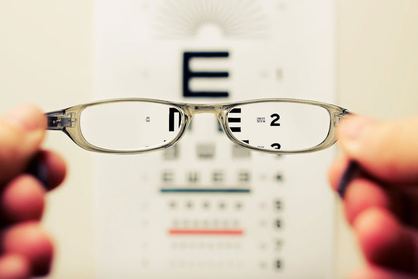
Snellen Eye Chart
- Purpose: Used for assessing visual acuity (sharpness of vision).
- Advantages: Simple and widely used for basic vision screening.
Visual Field Testing Equipment
- Purpose: Evaluate the full horizontal and vertical range of what an individual can see, detecting abnormalities or blind spots.
- Tools: Automated perimetry devices, confrontation visual field testing.
- Advantages: Crucial for conditions like glaucoma and neurological disorders affecting peripheral vision.
Refraction Tools (Phoropter, Autorefractor)
- Purpose: Measures refractive errors (nearsightedness, farsightedness, astigmatism).
- Tools: Phoropter (used in subjective refraction), Autorefractor (measures refractive errors objectively).
- Advantages: Essential for determining the need for glasses or contact lenses.
Optical Coherence Tomography (OCT)
- Purpose: Provides detailed cross-sectional images of the retina and optic nerve, aiding in the diagnosis and management of retinal conditions.
- Advantages: High-resolution imaging for assessing retinal thickness and detecting abnormalities.
Slit Lamp Biomicroscope
- Purpose: Allows detailed examination of the anterior segment of the eye, including the cornea, iris, and lens.
- Advantages: Essential for diagnosing corneal disorders, assessing contact lens fit, and evaluating eye structures.
iCare Tonometer
- Purpose: Measures intraocular pressure, important for glaucoma diagnosis and management.
- Advantages: Quick, contactless measurement, suitable for regular monitoring.

What is included in a standard eye test?
A standard eye test, also known as a comprehensive eye examination or routine eye checkup, typically includes a series of evaluations to assess various aspects of your eye health and vision. The exact components of an eye test may vary slightly based on the healthcare provider and the specific needs of the patient. However, a standard eye test often includes the following key elements:
Case History
- The eye care professional will start by gathering information about your general health, medications, family history of eye conditions, and any specific eye concerns or symptoms you may have.
Visual Acuity Testing
- Using an eye chart, usually the Snellen chart, the eye care professional measures your visual acuity at various distances to assess the sharpness of your vision.
Refraction Assessment
- The eye care professional uses a phoropter or autorefractor to determine if you have refractive errors such as nearsightedness, farsightedness, or astigmatism. This helps determine the prescription for glasses or contact lenses if needed.
Binocular Vision and Eye Movement Testing
- Evaluates how well your eyes work together and how effectively they move. This can include tests for eye alignment, depth perception, and tracking.
Ocular Health Examination
- The eye care professional examines the external and internal structures of the eye. This includes the eyelids, cornea, conjunctiva, iris, lens, and retina. Techniques such as slit lamp biomicroscopy and ophthalmoscopy may be used.
Intraocular Pressure Measurement
- The eye care professional may measure intraocular pressure to screen for glaucoma. This can be done with a tonometer, and more recently, contactless tonometers like the iCare tonometer.
Visual Field Testing (Optional)
- Assesses your peripheral vision and can help detect abnormalities or blind spots. This is important for conditions like glaucoma.
Retinal Imaging (Optional)
- Some eye care professionals may use imaging technologies such as fundus photography or optical coherence tomography (OCT) to capture detailed images of the retina and optic nerve.
Discussion of Findings and Recommendations
- The eye care professional will discuss the results of the tests, provide any necessary prescriptions, and make recommendations for further treatment or follow-up appointments.
Please remember that this is a general overview, and the specific tests performed may vary based on the practitioner's preferences and the patient's individual needs or concerns. Regular eye examinations are important for maintaining good eye health and detecting potential issues early. The frequency of eye exams may vary depending on factors such as age, overall health, and the presence of specific eye conditions. It's essential to follow the recommendations of your eye care professional.

What can opticians see behind the eye?
Opticians are not trained to directly observe the internal structures of the eye. Their primary role is to assess and address refractive errors (nearsightedness, farsightedness, astigmatism, and presbyopia) and fit eyeglasses or contact lenses based on prescriptions provided by optometrists or ophthalmologists.
Opticians typically work with prescriptions generated by eye care professionals, and their responsibilities include:
Interpreting Prescriptions
Opticians read and interpret prescriptions written by optometrists or ophthalmologists to determine the appropriate lenses for eyeglasses or contact lenses.
Fitting and Adjusting Eyewear
They assist customers in selecting frames or contact lenses and ensure that the eyewear fits comfortably. Opticians also adjust frames to achieve the best possible fit.
Lens Fabrication
Opticians may be involved in the fabrication and assembly of eyeglass lenses into frames, ensuring that the lenses meet the specifications provided in the prescription.
Educating Patients
Opticians educate patients about proper eyewear care, lens options, and any additional features or coatings that may benefit their vision.
If you need an examination of the internal structures of your eyes, such as the retina or optic nerve, or if you have specific eye health concerns, you should consult with an optometrist or ophthalmologist. These eye care professionals have the training and expertise to conduct comprehensive eye exams, diagnose eye conditions, and provide appropriate medical treatment or referrals if necessary.
Optometrists and ophthalmologists may use tools such as ophthalmoscopes, fundus cameras, optical coherence tomography (OCT), and other specialised equipment to examine the back of the eye and assess the health of the retina, optic nerve, and other structures. Regular eye exams with these professionals are essential for maintaining good eye health and detecting potential issues early.
What are urgent eye conditions?
Urgent eye conditions encompass a range of ocular issues that require prompt attention from an eye care professional to prevent potential vision loss or further complications. These conditions may include eye injuries, sudden vision changes, severe eye pain, persistent redness, flashes of light, or the sudden onset of floaters. Urgent eye conditions can arise from various causes, such as trauma, infections, glaucoma, retinal detachment, or corneal disorders. Seeking immediate medical attention is crucial in these situations to receive a timely and accurate diagnosis, initiate appropriate treatment, and prevent long-term damage to the eyes. Prompt intervention can often make a significant difference in preserving vision and addressing the underlying causes of urgent eye concerns. If experiencing any sudden or severe eye symptoms, individuals should seek urgent care from an optometrist, ophthalmologist, or visit the nearest emergency department.
Discover My-iClinic
Transform your vision and experience unparalleled eye care at My-iClinic. Our state-of-the-art facility is equipped with the latest technology for precision vision correction and comprehensive private eye care. Whether you're seeking advanced refractive surgery with our cutting-edge iTrace system, meticulous cataract surgery planning using the IOL Master, or detailed retinal imaging with Optical Coherence Tomography (OCT), My-iClinic is committed to providing the highest standard of care. Don't compromise on your vision – take the first step towards exceptional eye health. Schedule your appointment today and discover the My-iClinic difference, where innovation meets personalised care for a clearer and brighter future.
Find out more by Speaking to our team