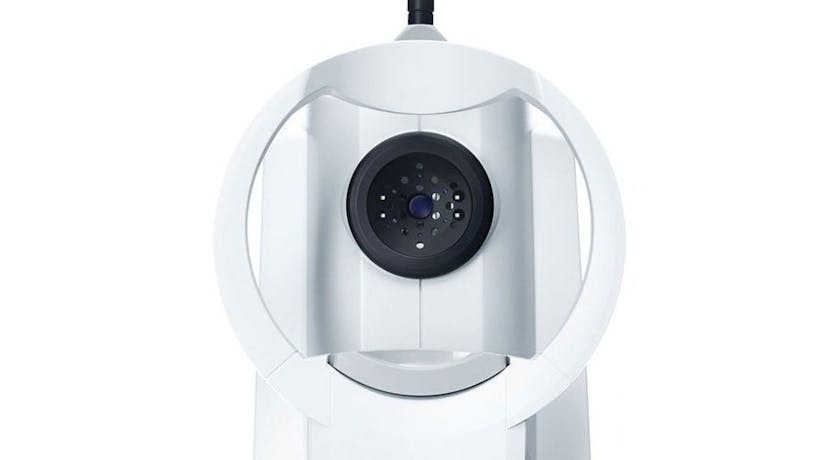
An epiretinal membrane, also known as a macular pucker or cellophane maculopathy, is a condition that affects the retina, the light-sensitive tissue at the back of the eye. This membrane is a thin, transparent layer of fibrous tissue that forms on the surface of the macula, which is the central part of the retina responsible for sharp, detailed vision. The development of an epiretinal membrane is often associated with the ageing process, where changes in the vitreous humor, the gel-like substance that fills the eye, can lead to the formation of scar tissue on the retinal surface. As the membrane contracts, it can cause wrinkling or distortion of the macula, impacting central vision and resulting in symptoms such as blurriness or mild distortion in the affected eye.
The severity of symptoms associated with an epiretinal membrane can vary, and not everyone with this condition experiences significant vision problems. However, in cases where visual impairment is notable, surgical intervention may be considered to remove the membrane and restore clearer vision. Regular eye examinations are crucial for detecting the presence of epiretinal membranes early on and determining the appropriate course of action to preserve and optimise visual function.
Is Epiretinal Membrane Serious?
While an epiretinal membrane (ERM) is not typically a life-threatening condition, it can have a significant impact on vision and visual quality. The seriousness of an epiretinal membrane depends on the severity of symptoms and how much it affects a person's daily life. Some individuals with ERMs may experience minimal or no visual disturbance, while others may notice blurriness, mild distortion, or decreased central vision in the affected eye.
In cases where the symptoms are mild, observation and regular monitoring may be recommended. However, if the membrane causes significant visual impairment or distortion, surgical intervention may be considered. Epiretinal membrane surgery, often performed through a procedure called vitrectomy, aims to remove the membrane and improve visual clarity.
It's essential to address symptoms associated with an epiretinal membrane promptly, as in some instances, it may lead to more serious complications such as macular edema or even a macular hole. Regular eye examinations and consultations with an eye care professional are crucial for monitoring the progression of the condition and determining the appropriate course of action to maintain optimal eye health and vision.

What Is Macular Edema?
Macular edema is a condition characterised by the accumulation of fluid in the macula, the central part of the retina responsible for detailed and sharp vision. This swelling can result from various underlying factors, such as diabetic retinopathy, macular degeneration, or vascular diseases. The increased fluid in the macula can lead to distorted or blurred vision and, if left untreated, may contribute to the deterioration of central vision. Macular edema is a serious concern, particularly in the context of chronic conditions like diabetes, where it is a common complication. Timely diagnosis and appropriate management, often involving treatments like anti-VEGF injections or laser therapy, are crucial to mitigate the impact of macular edema on visual function and prevent further vision loss. Regular eye examinations play a pivotal role in the early detection and intervention of this condition, optimising outcomes and preserving overall eye health.

What Is a Macular Hole?
A macular hole is a structural abnormality in the macula, the central part of the retina responsible for detailed and sharp vision. This condition involves the development of a small opening or defect in the macula, leading to a loss of tissue integrity. Macular holes can occur due to age-related changes, and certain risk factors include vitreous traction, eye trauma, or conditions like diabetic eye disease. The hallmark symptom of a macular hole is a sudden or gradual loss of central vision, often accompanied by distortion or a dark spot in the centre of the visual field. Prompt diagnosis through comprehensive eye examinations is crucial for determining the severity of the macular hole and guiding appropriate intervention. Surgical procedures, such as vitrectomy with the potential use of a gas bubble, are common treatments aimed at closing the hole and restoring visual function. Early detection and timely management are essential in optimising outcomes and preventing further deterioration of central vision associated with macular holes.

What Are The Symptoms Of Epiretinal Membrane?
ERM symptoms can vary in severity, and not everyone with an ERM will experience noticeable visual disturbances. However, common symptoms associated with an epiretinal membrane may include:
- Blurred Vision: Blurriness or haziness in central vision is a common symptom as the membrane affects the light entering the eye.
- Mild Distortion: Some individuals may perceive mild distortion in their vision, where straight lines may appear slightly curved or wavy.
- Reduced Central Vision: In more severe cases, there may be a noticeable decrease in central vision acuity, impacting activities like reading or recognising faces.
- Metamorphopsia: This term refers to a type of visual distortion where objects may appear larger, smaller, or distorted in shape.
- Difficulty with Fine Detail: Tasks requiring sharp vision and fine detail, such as reading or threading a needle, may become more challenging.
- Double Vision: Rarely, an epiretinal membrane may cause double vision, particularly if it affects the coordination of the eyes.
Furthermore, some individuals with an epiretinal membrane may not experience significant symptoms, and the condition may be detected during a routine eye examination. Regular eye check-ups are crucial for early detection and appropriate management of any visual issues associated with an epiretinal membrane. If you notice changes in your vision, especially in the central visual field, it's advisable to consult with an eye care professional for a thorough examination.
How To Fix Epiretinal Membrane
The treatment for an epiretinal membrane (ERM) depends on the severity of symptoms and how much it impacts a person's vision. Not everyone with an ERM requires treatment, especially if symptoms are mild and not significantly affecting daily activities. However, in cases where the membrane causes notable visual disturbances, surgical intervention may be considered. The primary surgical procedure for addressing an epiretinal membrane is called vitrectomy, and it involves the following steps:
Vitrectomy
During this surgery, the vitreous gel in the eye is removed, along with the epiretinal membrane. The surgeon may use microsurgical instruments to delicately peel off the membrane from the surface of the retina.
Gas or Silicone Oil
Following the removal of the vitreous and membrane, a gas bubble or silicone oil may be injected into the eye to help flatten the retina and facilitate the healing process. The choice between gas and silicone oil depends on the specifics of the case.
Positioning
Patients may be instructed to maintain a specific head position for a period after surgery to ensure proper placement of the gas bubble and optimal healing.
Recovery
Recovery times can vary, but patients are typically advised to avoid strenuous activities and certain head positions during the initial healing period.
While surgery can improve vision for many people with ERMs, the outcome is variable, and not everyone will experience a complete restoration of vision. Additionally, as with any surgery, there are potential risks and complications, and the decision to proceed with surgery should be made in consultation with an eye care professional who can assess individual circumstances and provide personalised recommendations. Regular follow-up appointments are crucial for monitoring the healing process and ensuring the best possible visual outcome.

FAQs
Q: What is an Epiretinal Membrane (ERM)?
A: An Epiretinal Membrane is a thin, transparent layer of fibrous tissue that forms on the surface of the macula, affecting central vision. It often develops due to changes in the vitreous humor within the eye.
Q: What Causes an Epiretinal Membrane?
A: The primary cause is age-related changes in the vitreous, leading to the formation of scar tissue on the retinal surface. Other factors may include eye trauma, inflammation, or vascular diseases.
Q: Are ERMs Common?
A: Yes, ERMs are relatively common, especially in older adults. Many people may have mild membranes without significant visual impact.
Q: What are the Symptoms of an Epiretinal Membrane?
A: Common symptoms include blurred vision, mild distortion, and reduced central vision. In more severe cases, objects may appear larger or smaller than they are.
Q: Can an Epiretinal Membrane Heal on its Own?
A: In some cases, mild membranes may not require treatment, and symptoms may remain stable or improve over time. However, surgical intervention may be considered for significant symptoms.
Q: How is an Epiretinal Membrane Diagnosed?
A: Diagnosis typically involves a comprehensive eye examination, including optical coherence tomography (OCT) imaging, which provides detailed images of the retina.
Q: What is Vitrectomy?
A: Vitrectomy is a surgical procedure used to remove the vitreous gel, along with the epiretinal membrane, from the eye. It is a common treatment for more severe cases of ERMs.
Q: Is Vitrectomy the Only Treatment Option?
A: While vitrectomy is a common and effective treatment, not all ERMs require surgery. Observation and monitoring may be recommended for mild cases.
Q: What is the Recovery Process After Vitrectomy?
A: Recovery times vary, but patients may need to avoid certain activities and maintain a specific head position for optimal healing. Follow-up appointments are crucial.
Q: Can Vision be Fully Restored After ERM Surgery?
A: The outcome varies, and while many people experience improved vision after surgery, complete restoration is not guaranteed. Regular follow-ups with an eye care professional are essential for monitoring progress.
Unlock Clarity and Renewed Vision with My-iClinic in London!
Are you experiencing blurred vision, mild distortion, or reduced central vision? These could be signs of an Epiretinal Membrane (ERM). At My-iClinic in London, our dedicated team of experienced eye care professionals specialises in providing advanced solutions for ERMs.
Benefit from our cutting-edge diagnostic technologies and personalised treatment plans designed to address your unique visual needs.
Why Choose My-iClinic?
- Expertise: Trust our skilled ophthalmologists with a wealth of experience in diagnosing and treating ERMs.
- State-of-the-art technology: Access the latest diagnostic tools and surgical techniques for precise and effective solutions.
- Patient-Centric Care: Experience compassionate care tailored to your specific condition and lifestyle.
Take the First Step Towards Clearer Vision!
Don't let an ERM compromise your eyesight. Schedule a consultation with My-iClinic today. Our team is committed to restoring clarity and optimising your visual health. Call us at 02084458877 or book your appointment. Your journey to clearer vision begins with My-iClinic in London!
Find out more by Speaking to our team


